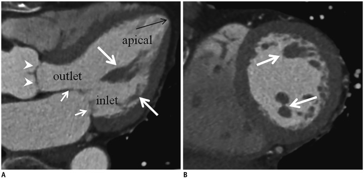
Fig. 1. Anatomy of left ventricle.
Three-chamber (A) and short-axis (B) MPR CCT images show mitral valve (short arrows), aortic valve (arrowheads), papillary muscles (thick arrows), and apical thinning (long arrow). Left ventricle is divided into three portions: inlet, apical, and outlet. Short-axis MPR image (B) is obtained at midventricular level. CCT = cardiac computed tomography, MPR = multiplanar reformat

