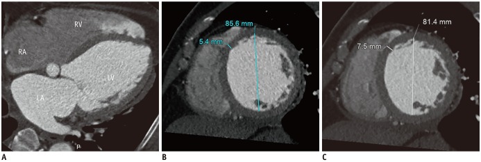
Fig. 12. DCM in 63-year-old man.
Four-chamber MPR image (A) obtained during end-diastole shows all cardiac chamber dilatation. Short-axis MPR CCT images obtained during end-systole (B) and end-diastole (C) show LV dilation, thinned myocardium (5 mm in thickness), and global severe hypokinesia. LV ejection fraction, end-diastolic, and end-systolic volumes were 12%, 137 mL, and 59 mL, respectively.

