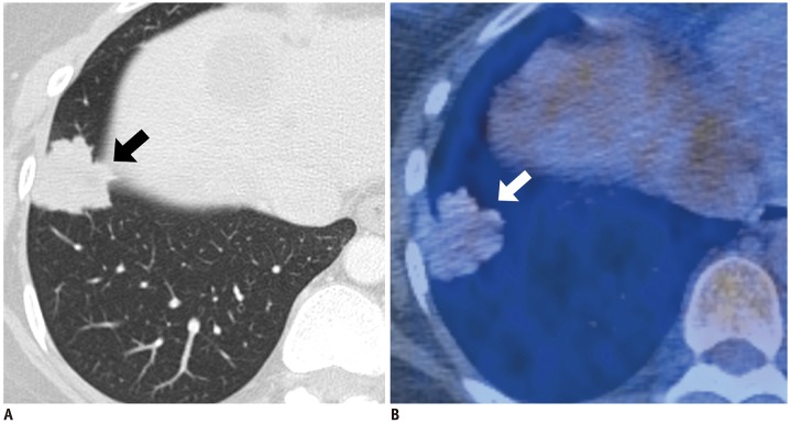
Fig. 2. IMA with positive M-M dissociation sign in 43-year-old woman.
A. Lung window image of transverse CT scan obtained at level of liver dome shows 30-mm-sized nodule with lobulated or spiculated margin (arrow) in right lower lobe (TDR = 1.34%). B. PET/CT image demonstrates scant 18F-FDG uptake (arrow) within tumor and with SUVmax of 2.2. FDG = fluorodeoxyglucose, M-M = morphologic-metabolic, SUVmax = maximum standardized uptake value, TDR = tumor shadow disappearance rate, 18F = fluorine-18

