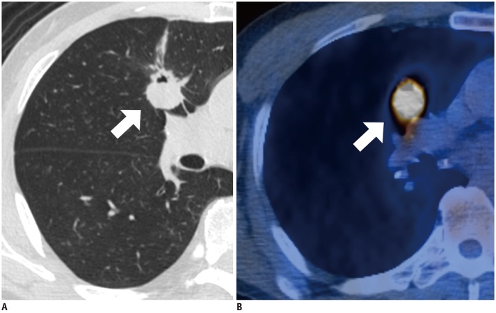
Fig. 4. Invasive non-mucinous adenocarcinoma with negative M-M dissociation sign in 60-year-old man.
A. Lung window image of transverse CT scan obtained at level of right bronchus intermedius shows 24-mm-sized lobulated and spiculated nodule (arrow) in right middle lobe (TDR = 35.31%). Additionally, note internal cavitation or bubble lucency within tumor. B. PET/CT image demonstrates hot 18F-FDG uptake (arrow) within tumor and with SUVmax of 12.9.

