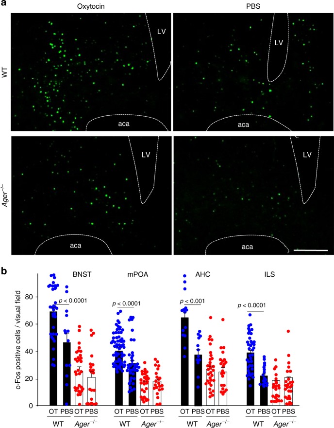
Fig. 6.
Reporter assay for oxytocin activity in the brain. a 60 min after sc administration of oxytocin (OT) (30 ng) or PBS, c-Fos positive nuclei were visualised in the bed nucleus of the stria terminalis (BNST) of WT and Ager−/− male mice (LV, lateral ventricle; aca, anterior commissure, anterior part) (Bar = 100 μm). b Densities of c-Fos positive nuclei in the BNST, medial preoptic area (mPOA), centre of the anterior hypothalamic area (AHC), and intermediate lateral septal nucleus (ILS) regions of WT and Ager−/− mice (n = 12–56)

