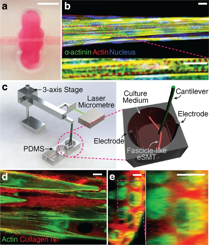
Figure 1.
Co-stimulation system and fascicle-inspired engineered skeletal muscle tissue (eSMT). (a,b) The fascicle-inspired eSMT formed a cylindrical shape with length of 6 mm and diameter of approximately 75 μm (a). The tissues were stained using immunofluorescence technique to visualize the striations of α-actinin, which is a marker of differentiation and contractility (b, reproduced from reference 40 with permission from the Mary Ann Liebert, Inc., New Rochelle, NY). Scale bars represent 5 mm in (a) and 10 μm in (b). (c) Schematic of the experimental setup used to apply coordinated electric and mechanical stimulation to the eSMT. The co-stimulation system consists of electrodes for applying the electric potential, a cantilever wire moved by a servomotor, and a laser micrometre to monitor the displacement of the cantilever. The eSMT is pulled sideways with the cantilever to stretch eSMT to the desired strain. Contractile force was quantified by measuring deformation of the cantilever whose bending stiffness is known. (d,e) Cross-sectional images of unstimulated eSMTs stained for collagen IV (red) and actin (green) in longitudinal (d) and transverse (e) directions. Scale bars represent 10 μm.

