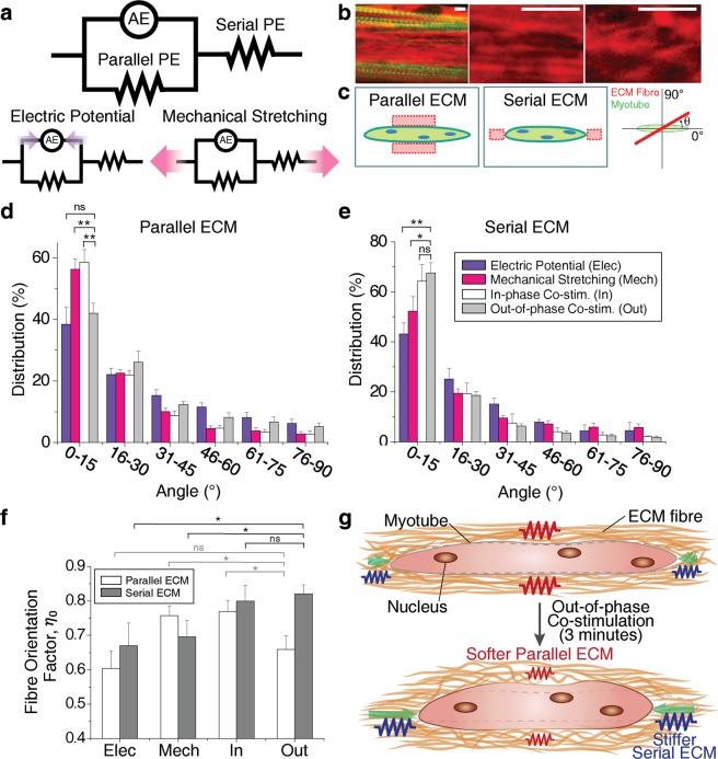
Figure 3.
Mechanistic model of eSMT force generation and transmission, and changes in the mechanical property induced by ECM remodelling. (a) Mechanistic model of eSMT consisting of an active element (AE), parallel passive element (Parallel PE), and serial passive element (Serial PE). (b) Images of collagen IV (red) and actin in the myotubes (green) of the eSMTs to measure the fibre orientation distribution of the ECM network (left). Images of well-aligned collagen fibres (middle) and less-aligned fibres (right). Scale bars represent 5 μm. (c) Schematics showing the regions of the parallel and serial ECMs relative to the myotubes, and ECM fibre orientation θ measured from the longitudinal direction of myotube. (d,e) Orientation distribution of collagen IV in the parallel (d) and serial (e) ECM regions. Most ECM fibres were aligned parallel to the longitudinal direction of the myotubes (0°) by pretension. SEM, n = 8, 12, 11, and 10 (parallel), n = 3, 7, 3, and 7 (serial). (f) Comparison of fibre orientation factors (ηo) to predict the elastic modulus of the ECM network (EL). The out-of-phase co-stimulation induced the highest elastic modulus (largest ηo) for the Serial PE and a low elastic modulus for the Parallel PE. SEM, n is the same as (d,e). *P < 0.05, **P < 0.01, and ns, not significant. (g) Schematic depicting the mechanism underlying the changes in performance induced by ECM remodelling following the application of the out-of-phase co-stimulation. The out-of-phase co-stimulation decreases the stiffness of the parallel ECM for less impedance on muscle contraction and increases the stiffness of the serial ECM to increase force transmission to the load.

