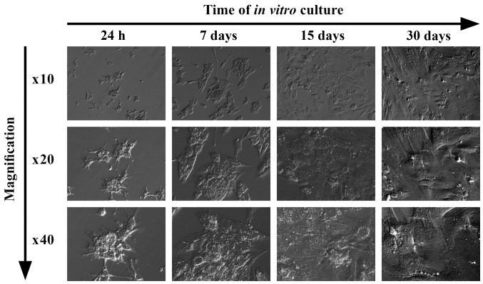
Figure 5.
Pictures of human ovarian granulosa cell cultures used for the experiment, presenting changes in their morphology following 24 h, and 7, 15 and 30 days of in vitro culture. All of the photographs were taken using an inverted microscope, under relief phase contrast (magnification, ×10, 20 and 40, as indicated).

