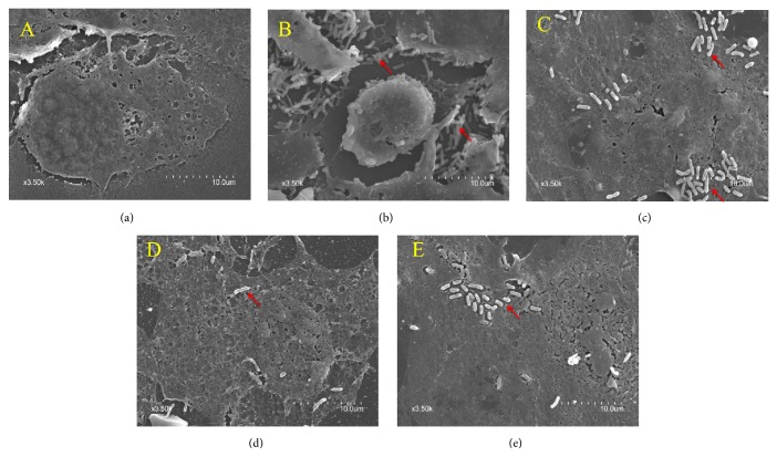
Figure 2.
Ultrastructural morphology of IPEC-J2 cells observed using scanning electron microscopy. Under normal conditions, the structure of IPEC-J2 cells was intact (a). Following ETEC infection, a large number of ETEC cells adhered to the surface of IPEC-J2 cells. ETEC damaged the structure of IPEC-J2 cells and caused shrinking of cellular morphology (b). Pretreatment with puerarin at 200 μg/mL (c), baicalin at 1 μg/mL (d), and berberine hydrochloride at 100 μg/mL (e) improved the structure and morphology of IPEC-J2 cells and decreased the number of adhered single bacteria ((a) control cells; (b) treatment with ETEC for 3 h; (c) pretreatment with puerarin 200 μg/mL + ETEC; (d) pretreatment with baicalin 1 μg/mL + ETEC; (e) pretreatment with berberine hydrochloride 100 μg/mL + ETEC; red arrows represent the single bacteria of ETEC).

