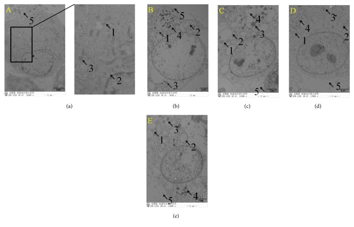
Figure 3.
Ultrastructural morphology of IPEC-J2 cells observed using transmission electron microscopy. Under normal conditions, the structure of IPEC-J2 cells was intact (a). Following ETEC infection, epithelial cell microvilli were shed from the cellular surface. Mitochondria increased in size and became more spherical, mitochondrial matrixes became shallower, and mitochondrial vacuolization was observed. Endoplasmic reticulum increased in size (b). In the puerarin (c), baicalin (d), and berberine pretreatment (e) groups, swollen mitochondria were also observed, the endoplasmic reticulum increased in size, the number of lysosomes increased, and digested fragments of bacteria were observed in the cytoplasm. Pretreatment with baicalin (d) significantly improved the structure of IPEC-J2 cells. 1 represents mitochondria, 2 represents endoplasmic reticulum, 3 represents lysosome, 4 represents bacterial fragments, and 5 represents epithelial cell microvilli ((a) control cells; (b) treatment with ETEC for 3 h; (c) pretreatment with puerarin 200 μg/mL + ETEC; (d) pretreatment with baicalin 1 μg/mL + ETEC; (e) pretreatment with berberine hydrochloride 100 μg/mL + ETEC).

