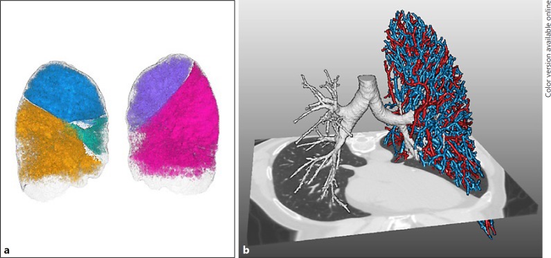
Fig. 1.
Example of development of quantitative HRCT software analysis (Thirona, Nijmegen, The Netherlands). a Rendering image of lobar volumes which is used for emphysema scores and air trapping and fissure analysis. b Rendering image of the bronchial tree (right lung) used for airway dimension analysis and pulmonary vasculature, used for vascular volume and lung perfusion.

