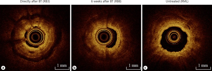
Fig. 3.
Optical coherence tomography (OCT) imaging immediately and 6 weeks after bronchial thermoplasty (BT) of BT-treated airways and of the untreated RML. OCT images captured in 1 procedure, in 1 patient, of the anterior segment of the right upper lobe (RB3) immediately after BT (a); the anterior basal segment (RB8) of the right lower lobe 6 weeks after treatment (b); and the BT-untreated right middle lobe (RML) (c).

