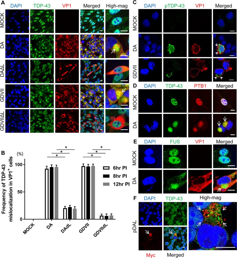
Fig 1. Mislocalization of TDP-43 in TMEV-infected cultured cells.
(A, D, E) Immunofluorescent staining of TDP-43, PTB1, and FUS in BHK-21 cells at 8 HPI. (A) TDP-43 is located in the nucleus of mock-infected (VP1-negative) cells. In DA and GDVII infections TDP-43 is depleted from the nucleus and mislocalizes to the cytoplasm of VP1-positive cells where it aggregates (arrows). In contrast, DAΔL and GDVIIΔL infections fail to induce TDP-43 mislocalization. (B) Frequency of TDP-43 mislocalization in TMEV-infected cells at different HPI. DA and GDVII infections induce mislocalization of TDP-43 in almost all VP1-positive cells that begins at least as early as 6 HPI and lasts for at least 12 HPI. In contrast, infection with TMEVΔL virus infrequently leads to TDP-43 mislocalization. (C) pTDP-43 is present in the cytoplasm of DA and GDVII-infected L929 cells (arrowheads). (D, E) PTB1 (D) and FUS (E) are mislocalized to the cytoplasm in DA infection (arrows). (F) 48hs after transfection with pDAL, TDP-43 is mislocalized and aggregates (arrows) in the cytoplasm of L-expressing BHK-21 cells (indicated by Myc positivity). Scale bars: 10 μm. *P < 0.001.

