Fig. 1.
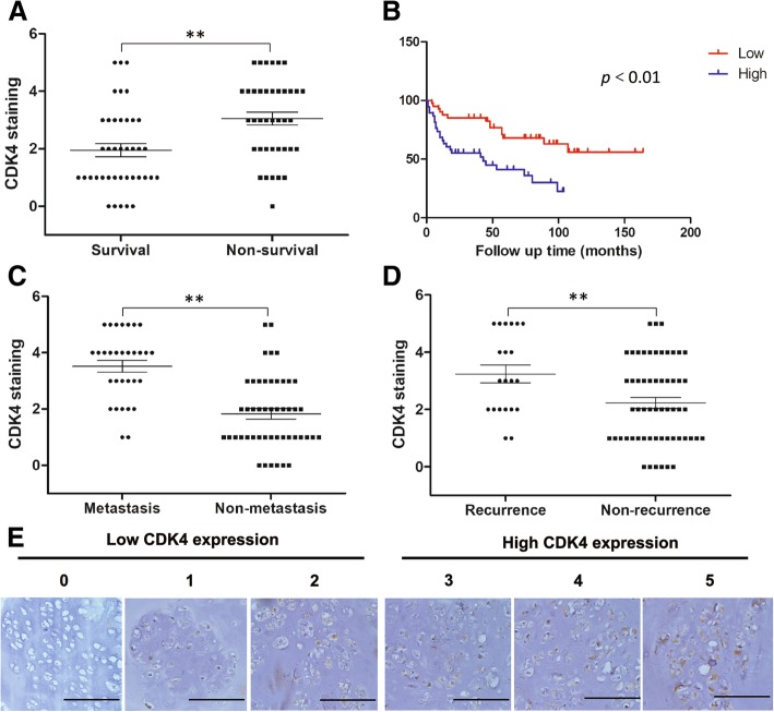
CDK4 expression levels are associated with the clinicopathological characteristics of chondrosarcoma patients. (a) Distribution of CDK4 staining scores in the chondrosarcoma tissue samples from surviving and non-surviving patients. (b) Kaplan-Meier survival curve of sarcoma patients with high staining (≥ 3) or low staining (< 3) for CDK4. Distribution of CDK4 staining scores in the chondrosarcoma tissue samples from patients with and without metastasis (c), patients with and without recurrence (d). ** means P < 0.01 compared with the former group. (e) Representative images of different immunohistochemical staining intensities of CDK4 (Original magnification 200X, scale bar =100 μm). On the basis of the percentage of cells with positive nuclear staining, CDK4 staining patterns were categorized into 6 groups: 0, no nuclear staining; 1+, < 10% positive cells; 2+, 10–25% positive cells; 3+, 26–50% positive cells; 4+, 51–75% positive cells; 5+, > 75% positive cells. Tumors with a staining score of ≥3 were designated as high CDK4 expression and < 3 were designated as low CDK4 expression

