Fig. 5.
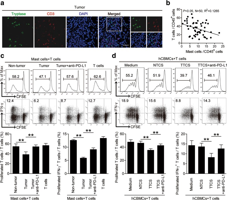
Tumor-infiltrating and tumor-conditioned mast cells suppress T cell immunity through PD-L1. a Representative analysis of tryptase+ mast cell (green) and CD3+ T cell (red) interactions in tumor tissues of GC patients by immunofluorescence. Scale bars: 20 μm. b The correlations between mast cells and T cells in human GC tumors were analyzed. Results were expressed as percentage of mast cells and T cells in CD45+ cells in tumor tissues of GC patients. c CFSE (carboxyfluorescein succinimidyl amino ester)-labeled peripheral CD3+ T cells of patients with GC were co-cultured for 5 days with autologous mast cells from non-tumor or tumor tissues with or without anti-PD-L1 antibody. Representative data and statistical analysis of T cell proliferation and IFN-γ production were shown (n = 5). d CFSE-labeled peripheral CD3+ T cells of donors were co-cultured for 5 days with autologous TTCS-, or NTCS-conditioned hCBMCs with or without anti-PD-L1 antibody. Representative data and statistical analysis of T cell proliferation and IFN-γ production were shown (n = 5). Each dot in panel b represents 1 patient. **, P < 0.01 for groups connected by horizontal lines. hCBMCs, human umbilical cord blood-derived cultured mast cells

