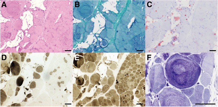
Fig. 1.
Muscular pathological findings (bar = 100 μm). a & b. H&E stain (a) and MGT stain (b): fibrosis, adipose tissue infiltration, rounding of muscle fibers, increased variability of fiber diameter, myonecrosis, and few regenerating fibers were seen. c. ORO stain: predominant adipose tissue infiltration was observed. d & e. ATPase stain(d: pH = 4.3, E: pH = 10.4): type-I and type-II fibers were affected equally with fiber type grouping. f. NADH stain: Disorganization of myofibril arrangement was noted on NADH stain

