
Fig. 5. Photomicrographs of lung tissue from MCA/GMAW-MS fume-exposed mice.
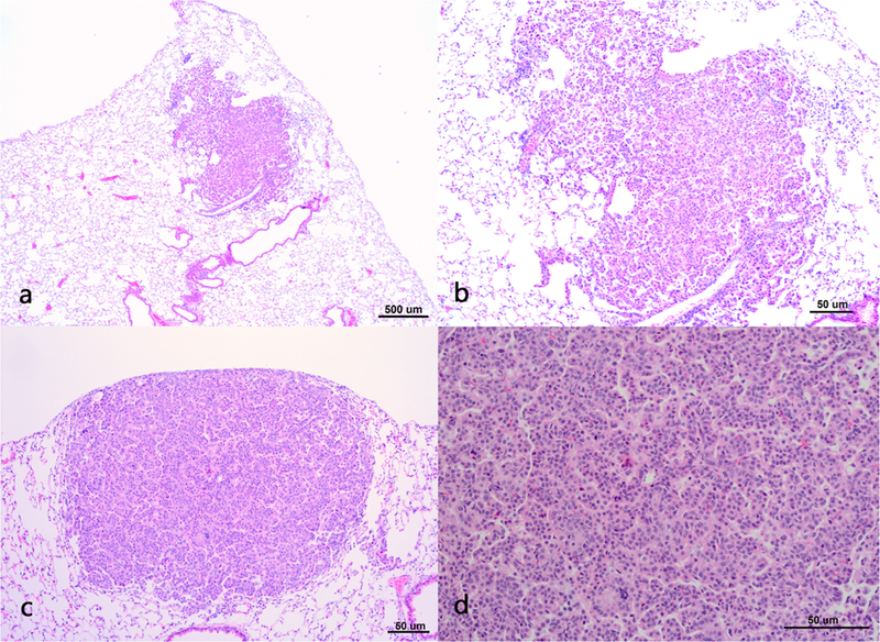
Panel A- moderate bronchioloalveolar hyperplasia, 4x magnification. Panel B – moderate bronchioloalveolar hyperplasia,10x magnification. Panel C – bronchioloalveolar adenoma as a solid discrete mass, 10x magnification. Panel D - bronchioloalveolar adenoma, 20x magnification.