Fig. 1.

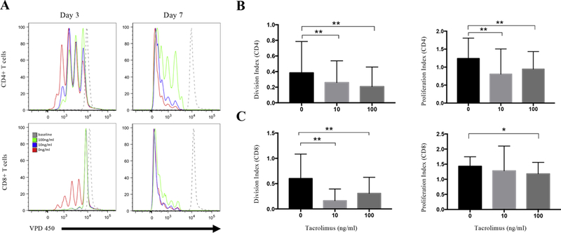
Tacrolimus decreases the proliferation of CD4+ and CD8+ T cells. PBMCs were isolated from 16 MuSK-MG patients, labeled with VPD450, and incubated with 1ug/ml anti-CD3 and 1ug/ml CD28 Abs in the absence or presence of 10ng/ml, 100ng/ml tacrolimus. (A) Comparison of CD4+ and CD8+ T cell proliferation in the presence of tacrolimus. The upper line shows CD4+ T cells proliferation after 3 days (left) and 7 days (right) of culture in the absence of tacrolimus (red), 10ng/ml tacrolimus (blue),100ng/ml tacrolimus (green), and the baseline was set on day 0 (grey, dashed line). The bottom histograms show CD8+ T cells proliferation after 3 days (left) and 7 days (right) of culture in the absence of tacrolimus (red), 10ng/ml tacrolimus (blue),100ng/ml tacrolimus (green), and the baseline was set on day 0 (grey, dashed line). (B) The division index (left) and proliferation index (right) in CD4+ T cells after 3 days of culture. (C) The division index (left) and proliferation index (right) in CD8+ T cells after 3 days of culture. *, p≤0.05; **, p≤0.01.
