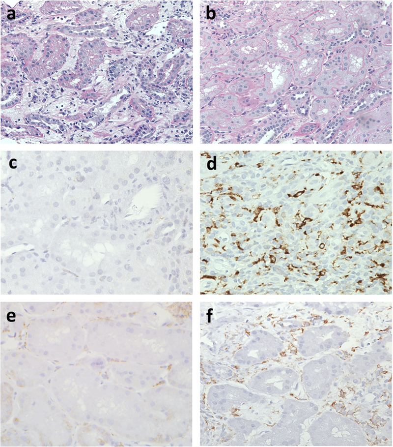
Fig. 4.

CD163 histopathology (brown stain) of representative allografts. a Normal kidney devoid of inflammation and edema. The non-atrophic tubules are back to back and the interstitium is barely visualized at ×200 magnification. b An acutely rejecting kidney shows interstitial expansion by edema characterized by loose myxoid stroma. Interstitial inflammation and tubulitis typical of T cell-mediated rejection are seen at ×200 magnification. c A non-rejecting allograft with no evidence of macrophage infiltration (negative CD163 stain) at ×400 magnification. d A non-rejecting allograft with parenchymal scarring possibly due to reflux nephropathy showing numerous CD163-positive macrophages at ×400 magnification. e An allograft undergoing rejection with minimal macrophage infiltration at ×400 magnification. f An allograft with histological features of both tubulointerstitial T cell-mediated and antibody-mediated acute rejection showing numerous CD163-positive macrophages within the interstitium and peritubular capillaries at ×400 magnification.
