. 2019 Feb 4;8:e42692. doi: 10.7554/eLife.42692

Figure 3. Effects of mutating DIP-α on the terminal branching of α-leg MNs.

(A) Left Column: Proximal-Distal (P–D) oriented Fe and Ti T1 adult leg segments depicting axon muscle-targeting of three DIP-α expressing leg MNs labeled by DIP-α-A8-Gal4(86Fa)>20XUAS-6XGFP (green) (Figure 3—figure supplement 1D) (See Materials and methods). Muscles are labeled using Mef2-QF > 10XQUAS-6XmCherry (red); Grey; cuticle. MNs are named according to the muscle target (αFe-ltm, αTi-ltm, and αTi-tadm) (Soler et al., 2004). Right Columns: Transverse sections of Fe and Ti leg segments at specific locations along the P-D axis, corresponding to the numbered white dotted lines on the left, depicting terminal branching (green arrows) on the Fe and Tiltms (encircled by white dotted lines) and tadm. (scale bar: 50 μm). (B) Terminal branching of the T1 α-leg MNs labeled by DIP-α-A8-Gal4(86Fa)>20XUAS-6XGFP in wild type (WT), DIP-α mutant and rescue contexts. Left; T1 legs; Right; Fe and Ti leg segments (axons; green (WT/rescue) or white (mutant), cuticle; grey). Absence of terminal branching of the α-ltm MNs in the DIP-α mutant T1 leg is indicated by white dotted circles; White arrowheads demarcate axons reaching the vicinity of their muscle targets (refer to Figure 3—figure supplement 1D). (scale bar: 50 μm). (C) Intermediate terminal branching defects in T1 legs displayed by αTi-ltm and αTi-tadm in DIP-α mutants. Single cell labeling of αTi-ltm and αTi-tadm terminal branches in the T1 proximal Ti is shown in WT (green) and DIP-α mutant (white). (scale bar: 50 μm). (D) Quantification of mutant phenotypes (αFe-ltm, light green; αTi-ltm, medium green; and αTi-tadm, dark green) in WT (N = 20), mutant (diagonal lines) and rescue contexts (N = 7 to 20) using a DIP-α null, chromosomal deficiency and MiMIC-T2A-Gal4/QF as indicated. Statistical significance was determined using Fisher’s exact test: *p<0.05; **p<0.01; ***p<0.001 (E) Quantification of number of branches on αTi-ltm and αTi-tadm single-cell samples in WT and DIP-α mutant contexts using genotypes indicated in Figure 1C. Statistical significance was determined using a two-tailed unpaired t-test for αTi-ltm samples, where error bars represent mean ± SD and a Mann-Whitney U test for αTi-tadm samples, where error bars represent median ± interquartile ranges. ***p<0.001 (F) Ectopic expression of DIP-α in LinB/24 leg MNs targeting the Coxa, Trochanter and Distal Fe using OK371-Gal4 MARCM (Top) or an enhancer trap hkb-Gal4 (Bottom) which also labels an additional leg MN targeting the distal Fe (white arrowhead). Normal axon targeting of LinB/24 leg MNs (white) is shown on the left without any terminal branching at the Fe-ltm. However, in a rare case (N = 1/9), ectopic expression of DIP-α using hkb-Gal4 in a DIP-α mutant background caused ectopic branching at the Fe-ltm (white arrowhead within magnified inset). (scale bar: 50 μm).

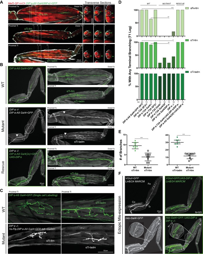
Figure 3.

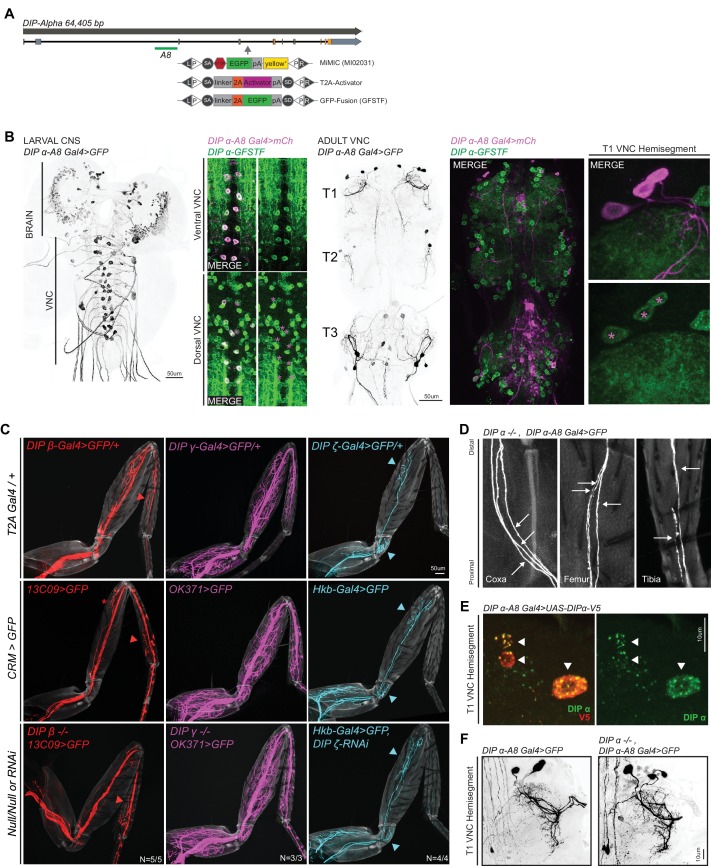
Figure 3—figure supplement 1. Characterization of DIP-α and other DIP mutants.

Figure 3—figure supplement 1.

(A) Genomic locus of DIP-α showing introns (black line), non-coding exons (grey rectangles) and coding exons (orange rectangles). The location of the DIP-α-A8 enhancer is denoted by a green bar (see Materials and methods). The location of the MiMIC insertion (MI02031) in a coding intron is denoted by a grey arrow. Using recombination mediated cassette exchange (RMCE), MI02031 has been independently swapped into a T2A-binary activator (Gal4/QF) that acts as a ‘gene trap’ and a GFP protein fusion (GFSTF) that acts as a ‘protein trap’ (see Materials and methods) (Venken et al., 2011). (B) Expression of DIP-α-A8-Gal4 > 20XUAS-6XGFP in the larval CNS and adult VNC (black, scale bar: 50 μm). DIP-α-A8-Gal4 is expressed in two segmentally repeated rows of cells along the length of the larval VNC (ventral and dorsal) and in the adult VNC, DIP-α-A8-Gal4 is expressed specifically in three leg MNs in each hemisegment of the thoracic ganglia, in addition to 6–8 cells in the abdominal ganglia (in between the T3 hemisegments). Co-expression of DIP-α-A8-Gal4 > 20XUAS-6XmCherry (Magenta) and DIP-α-GFSTF (detected by anti-GFP – see Materials and methods) in the ventral and dorsal larval VNC as well as the T1 hemisegment of the adult VNC, shows that the DIP-α-A8-Gal4+ cells (marked by magenta asterisks in the dorsal larval VNC and adult T1 VNC hemisegment) express DIP-α. (C) Comparisons of terminal branching of DIP-expressing T1 leg MNs (MiMIC-T2A-Gal4/+; Top Row) in WT (Enhancer-Gal4 >20XUAS-6XGFP; Middle Row) and mutant contexts (bottom row); DIP-β (red), DIP-γ (magenta) or DIP-ζ (cyan) (grey, cuticle). 13C09-Gal4 labels a DIP-β-expressing leg MN in the distal Ti (red asterisk marks a leg MN in the distal Fe that is inconsistently labeled even in WT animals); OK371-Gal4 labels the DIP-γ-expressing leg MNs targeting the majority of adult leg muscles; hkb-Gal4 labels DIP-ζ-expressing leg MNs in the Tr and distal Fe. DIP-expressing leg MN terminal branches that are consistently labeled by the Enhancer-Gal4 (colored arrowheads) are maintained in the mutant contexts. (scale bar: 50 μm). (D) Magnified images of T1 leg segments showing individual axons (white arrows) of the DIP-α mutant leg MNs from Figure 3B. Three axons enter the Coxa (left) and proximal Fe (middle) and two axons enter the proximal Ti (right) (grey, cuticle). (E) Expression of DIP-α in the T1 adult VNC hemisegment of DIP-α-A8-Gal4 > UAS-DIP-α-V5 animals detected by anti-V5 (red) and anti-DIP-α (green) (see Materials and methods). Anti-DIP-α worked to detect DIP-α protein levels only in the cell bodies when overexpressed. White arrowheads denote DIP-α-A8+ leg MNs. (scale bar: 10 μm). (F) No major abberrations were detected in the leg MN cell bodies and dendritic projections (Black) of the α-leg MNs by comparing WT (Left) and mutant (Right) adult T1 VNC hemisegments. (scale bar: 10 μm).
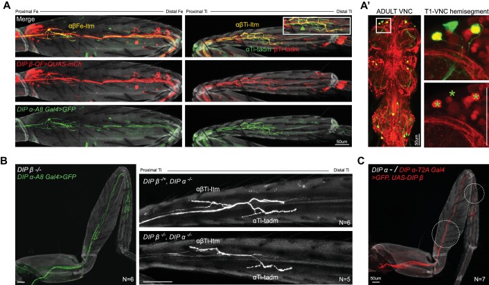
Figure 3—figure supplement 2. Co-expression and phenotypic analysis of DIP-α and DIP-β in adult leg MNs.

Figure 3—figure supplement 2.

(A) Expression of DIP-β in the α-ltm-leg MNs is confirmed by co-expression of DIP-β-T2A-QF > 10XQUAS-6XmCherry (red) along with DIP-α-A8-Gal4 > 20XUAS-6XGFP (green). (A) In the Fe (left), DIP-β is expressed in the DIP-α-expressing MN innervating the Fe-ltm (denoted as αβFe-ltm in yellow) while DIP-β alone is expressed in leg MNs innervating the distal Fe. In the Ti (right), DIP-β is expressed in the DIP-α-expressing MN innervating the Ti-ltm (denoted as αβTi-ltm in yellow) as well an additional proximal Ti-ltm innervating neuron that does not express DIP-α. Both DIP-α and DIP-β are independently expressed in two tadm-targeting leg MNs (denoted as αTi-tadm in green and βTi-tadm in red) whose terminal branches are closely associated with one another; Magnified inset; green arrowhead points to the αTi-tadm GFP +axon that does not express mCherry. (grey; cuticle). In the adult VNC (A’), co-expression of DIP-β-T2A-QF > 10XQUAS-6XmCherry (Red) with DIP-α-A8-Gal4 > 20XUAS-6XGFP (green) confirms that DIP-β is expressed in only two of three DIP-α-expressing leg MNs as seen in the magnified image of the T1 VNC hemisegment (green asterisks demarcate the locations of the DIP-α-A8-Gal4+ leg MN cell bodies). (scale bar: 50 μm). (B) Left: Terminal branching of the T1 α-leg MNs is unaffected in a DIP-β mutant (green, axons; grey, cuticle). Right: T1 Ti leg segments of a DIP-α homozygous and DIP-β heterozygous mutant (top) in comparison to a DIP-α and DIP-β homozygous double mutant (bottom) showing no increase in the severity of the terminal branching defect seen in αβTi-ltm (Figure 3.C). (white, axons; grey, cuticle). (scale bar: 50 μm). (C) DIP-β cannot rescue the DIP-α mutant branching defect when reintroduced in the mutant DIP-α leg MNs using DIP-α-T2A-Gal4 > UAS-DIP-β. (red, axons; grey, cuticle). Note that mutant αTi-tadm branching was not affected by ectopically expressing UAS-DIP-β. (scale bar: 50 μm).