. 2019 Feb 4;8:e42692. doi: 10.7554/eLife.42692

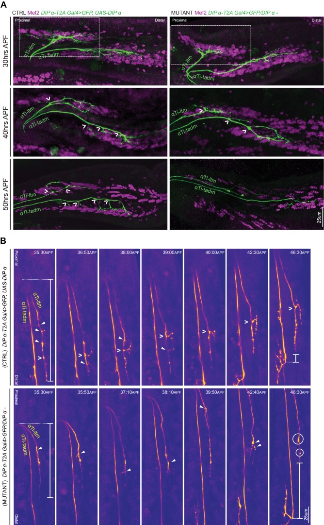
Figure 6. DIP-α is required for terminal axon lengthening and branching 30 to 45 hr APF.

(A) Terminal axon branching of control (left) and DIP-α mutant (right) αTi-ltm and αTi-tadm leg MNs at 30 hr (top), 40 hr (middle) and 50 hr (bottom) APF using DIP-α-T2A-Gal4 > UAS-DIP-α and DIP-α-T2A-Gal4/DIP-α, respectively. Axons are labeled using DIP-α-T2A-Gal4 > 20XUAS-6XGFP (green) and muscles are labeled with antibody against Mef2 (magenta). White arrowheads demarcate branch points along the axon terminal. At 50 hr APF, mutant αTi-ltm axons lack a prominent contralateral branch and mutant αTi-tadm axons lack four contralateral branches and retain the distal-most branch. White-dotted box denotes magnified inset in Figure 5—figure supplement 1A (scale bar: 25 μm). (B) Snapshots from time-lapse videos (Video 4, Video 5) comparing control (top) and mutant (bottom) αTi-ltm and αTi-tadm axons between ~35 hr and 45 hr APF (time-stamp is located on the top-right corner of each snapshot). Axons are labeled using DIP-α-T2A-Gal4 > 20XUAS-6XGFP (yellow). White open arrowheads demarcate the contralateral branch point on the αTi-ltm axon in the control sample while filled white arrowheads demarcate assorted dynamic filopodial projections along the αTi-ltm axon in both control and mutant samples. The distal-most tip of the αTi-ltm axon is more proximally located in the mutant sample compared to the control at ~35 hr APF (far left), as measured from the axon ‘bend’ at the joint between the distal Femur and proximal Tibia (denoted by white vertical bars) as well as at ~45 hr APF (far right), as measured from the distal most branch of αTi-tadm (denoted by white vertical bars). White circles demarcate globular punctate structures that form on the mutant αTi-ltm axon by ~45 hr APF (far right). (scale bar: 25 μm).

Figure 6.

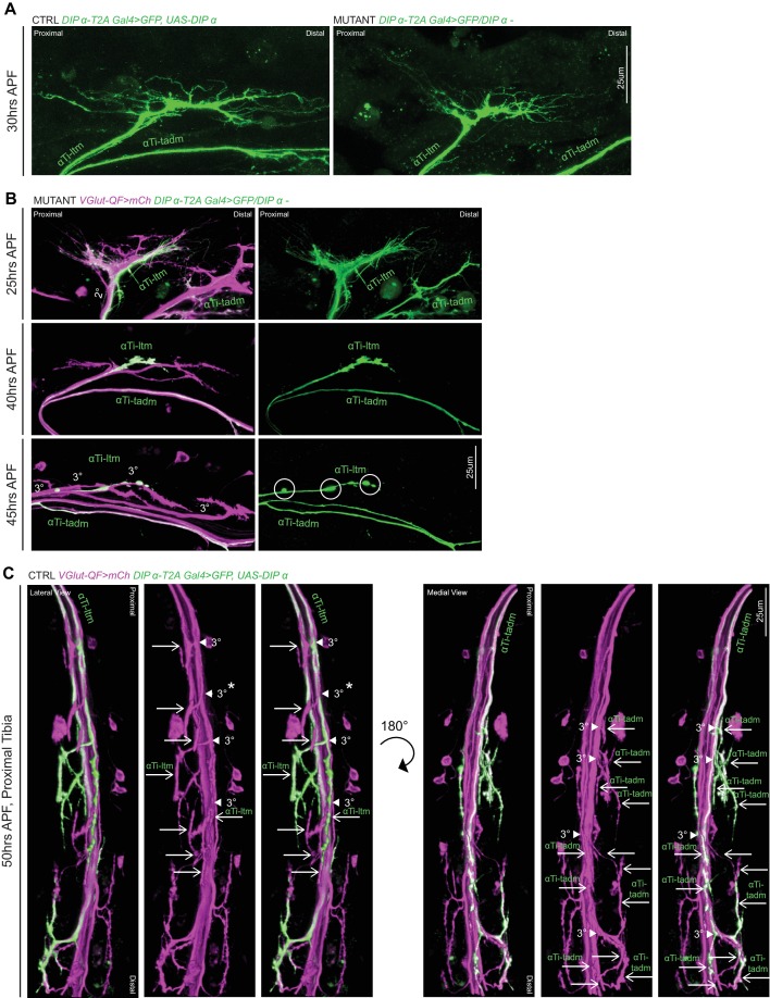
Figure 6—figure supplement 1. DIP-α is Required for terminal axon lengthening and branching 30 to 45 hr APF.

Figure 6—figure supplement 1.

(A) Magnified inset from Figure 6A showing decreased αTi-ltm terminal axon branching in a DIP-α mutant (right) compared to the control (left) at 30 hr APF. Axons are labeled using DIP-α-T2A-Gal4 > 20XUAS-6XGFP (green) (scale bar: 25 μm). (B) 3D projections of axon terminals of Ti-ltm targeting leg MNs, including αTi-ltm, in a DIP-α mutant background at 25 hr (top), 40 hr (middle) and 45 hr (bottom) APF. Leg MNs are labeled using VGlut-QF >10XQUAS-6xmCherry (magenta) and αTi-ltm is labeled using DIP-α-T2A-Gal4 > 20XUAS-6XGFP (green) (left, merge; right, αTi-ltm). At 25 hr APF, DIP-α mutant αTi-ltm axon terminals are located within their secondary axon bundles (2°) and generate filopodial projections. Between 40–45 hr APF, although mutant αTi-ltm axons sort into tertiary axon bundles (3°), they fail to generate terminal branches. White circles denote globular, punctate structures that form on the mutant αTi-ltm axon by ~45 hr APF. (scale bar: 25 μm). (C) 3D projections of axon terminals of Ti targeting leg MNs, including αTi-ltm (lateral view) and αTi-tadm (medial view), in a control background at 50 hr APF. Leg MNs are labeled using VGlut-QF >10XQUAS-6xmCherry (magenta) and α-leg MNs are labeled using DIP-α-T2A-Gal4 > 20XUAS-6XGFP (green)(left-unlabeled, merge; middle-labeled, VGlut; right-labeled, merge). White arrowheads point to tertiary bundles of MNs targeting the Ti-ltm and tadm and white arrows point to distinct terminal branches. In total there are four tertiary bundles targeting the Ti-ltm (one of which does not belong to the LinA/15 leg MN lineage – white asterix) composed of eight terminal branches, two of which belong to αTi-ltm. In the portion of the tadm pictured here, there are four tertiary bundles composed of approximately thirteen distinct terminal branches, of which nine belong to αTi-tadm.(scale bar: 25 μm).