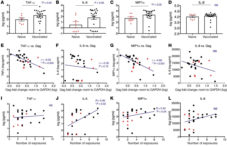
Figure 7. Enhanced expression of trained-immunity markers was inversely correlated with SHIV Gag expression ex vivo and positively with number of viral exposures in vivo.
PBMC samples collected from the vaccinated (1 week after the last boost) and naive animals were thawed, and the monocytes were enriched by 3 washes with warm PBS to remove the suspended cells after 2 hours of adherence to 48-well plates. The adherent monocyte-enriched cell populations were then cocultured with SHIV (1:100) for 18 hours. The supernatant was collected and the production of TNF-α, IL-6 (both as classical trained-immunity markers), MIP1α (SHIV coreceptor agonist), and IL-8 (negative control unrelated to trained immunity) were measured. The cells were further cultured for 2 more days before the expression levels of SHIV Gag in the cells of each sample were measured by qPCR. (A–D) The protein levels of TNF-α, IL-6, MIP1α, and IL-8 were compared between the naive and vaccinated animals. Mann-Whitney test was used for comparisons. Mean ± SEM are shown. (E–H) The cellular expression of SHIV Gag RNA was inversely correlated with TNF-α, IL-6 (trend), and MIP1α (but not with IL-8) production in the supernatant. Spearman’s r and P values of the correlations are indicated. (I–L) IL-6 and MIP1α (but not TNF-α or IL-8) were positively correlated with the number of viral exposures required for the animals to become infected in vivo. Jonckheere-Terpstra test was used to calculate the r and P values. Naive n = 7, shown in red triangles; vaccinated n = 21, shown in black dots. NS, not significant.

