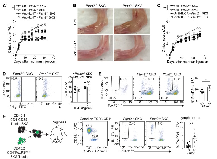
Figure 6. IL-6 promotes arthritis and Treg conversion in Ptpn2-haploinsufficient mice.
(A) Clinical score of male SKG mice after treatment with anti–IL-17A antibodies once weekly (100 μg i.p.; Ptpn2+/+, n = 5; Ptpn2+/–, n = 4) or control (Ptpn2+/+, n = 8; Ptpn2+/–, n = 8) during mannan-induced arthritis. (B) Representative images of ankles of 4 individual Ptpn2+/+ and Ptpn2+/– SKG mice treated with anti–IL-17A or control. (C) Clinical scores of male Ptpn2+/+ and Ptpn2+/– SKG mice treated with anti–IL-6R antibody once weekly (200 μg i.p.; Ptpn2+/+, n = 3; Ptpn2+/–, n = 3) or control (Ptpn2+/+, n = 5; Ptpn2+/–, n = 5) during mannan-induced arthritis. (D) In vitro differentiation of Th17 cells from naive Ptpn2+/+ (n = 4) and Ptpn2+/– (n = 5) SKG CD4+ T cells. (E) Conversion of flow-sorted Ptpn2+/+ (n = 4) and Ptpn2+/– (n = 3) SKG Tregs (CD4+FoxP3eGFP+) into IL-17–producing exTregs (IL-17A+FoxP3eGFP–) after 72 hours of stimulation with IL-6 (50 ng/ml) and anti-CD3/CD28–coated beads in vitro. (F) Cotransfer of CD45.1 SKG CD4+CD25– T cells with CD45.2 SKG Tregs to Rag2-KO mice. Transferred CD45.2 Ptpn2+/+ (n = 7) and Ptpn2+/– (n = 9) SKG Tregs were analyzed in lymph nodes of arthritic mice. Compiled data from at least 2 independent experiments are shown. Each symbol in D–F represents an individual mouse. Arthritis severity was quantified using the area under the curve. Bars represent mean ± SEM. *P < 0.05, **P < 0.01 by Mann-Whitney (A and C) or unpaired t test (E and F).

