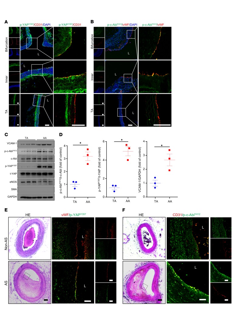
Figure 6. p-c-AblY412 and p-YAPY357 were highly expressed in ECs of atheroprone regions in both mouse and human.
(A and B) Aortas from 6- to 8-week-old Apoe–/– mice underwent immunofluorescence staining for indicated proteins. Bifurcation, bifurcation of aortic arch; Inner, inner curvature of aortic arch; TA, thoracic aorta; L, lumen. Represent images are shown, n = 6. Scale bars: 80 μm. (C and D) Protein was extracted from the aortic arch (AA) and TA of 8-week-old Apoe–/– mice. (C) Western blot analysis of expression of VCAM-1, p-YAPY357, t-YAP, p-c-AblY412, c-Abl, and GAPDH in the tissue lysates of AA and TA intima. (D) Quantification of protein expression in panel C. Data are mean ± SEM, *P < 0.05 (Student’s t test). Protein extracts of intima from 3 mice were pooled as 1 sample, n = 3. (E and F) Human atherosclerotic vessels were divided into atherosclerosis (AS) and non-AS groups. The vessels underwent HE staining and immunofluorescence staining for indicated proteins. Black frame indicates area magnified in immunofluorescence images. L, lumen. Representative images are shown, n = 10. Scale bars: 1000 μm (HE), 200 μm (immunofluorescence).

