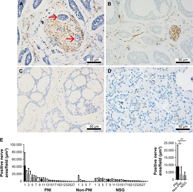
Figure 1.
Sympathetic innervation correlated positively with PNI in SACC tissues.
Notes: (A) Sympathetic nerve fibers were invaded by tumor cells in SACC tissues with PNI (invaded tumor cells are indicated with red arrowheads). (B) Fine sympathetic nerve fibers were mainly distributed in the stroma of SACC tissues without PNI. (C) Negative sympathetic innervation in SACC tissues without PNI. (D) Tiny sympathetic nerve fibers were sparsely scattered in NSG tissues. (E) Quantitative comparison of the sympathetic innervation areas in SACC tissues with PNI, SACC tissues without PNI, and NSG tissues. Left, quantification of sympathetic innervation areas in PNI SACC tissues (n=27), non-PNI SACC tissues (n=7), and NSG tissues (n=28). Each bar represents the average sympathetic innervation areas of three to five fields obtained from SACC or NSG tissue. Right, average sympathetic innervation areas in PNI SACC tissues, non-PNI SACC tissues, and NSG tissues. Original magnification, 400×; scale bar =50 μm. *P<0.05, **P<0.01. The error bars indicate the SD.
Abbreviations: NSG, normal salivary gland; PNI, perineural invasion; SACC, salivary adenoid cystic carcinoma.

