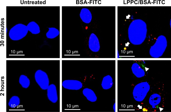
Figure 5.
Localization of LPPC/BSA-FITC complexes in HepG2 cells.
Notes: HepG2 cells were untreated (left panels) or treated with BSA-FITC (central panels) or LPPC/BSA-FITC complexes (right panels). The lysosomes and nuclei of the cells were individually stained red and blue, respectively. The cells were observed and imaged by confocal microscopy. Localization of BSA-FITC and lysosomes is indicated by white arrows. The escape of BSA-FITC from the lysosome (at 2 hours postincubation) is indicated by a white arrowhead. Representative images of three independent experiments are shown.
Abbreviations: FITC, fluorescein isothiocyanate; LPPC, liposomes containing polyethylenimine and polyethylene glycol complex.

