Figure 6.
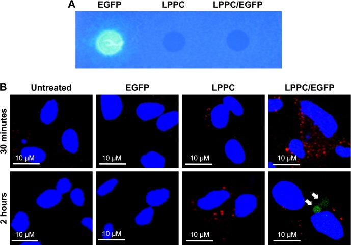
Escape of intracellularly delivered EGFP from LPPC.
Notes: (A) The fluorescence of EGFP cannot be measured, when it is adsorbed to LPPC (right dot), while free EGFP is capable of emitting fluorescence (left dot). (B) HepG2 cells were untreated or treated with free EGFP, empty LPPC, or LPPC/EGFP complexes for different amounts of time. The lysosome (in red color) and nucleus (in blue color) were stained after incubation. The cells were then observed and imaged by confocal microscopy. EGFP molecules dissociated from LPPC are indicated by white arrows. Representative images of three independent experiments are shown.
Abbreviations: EGFP, enhanced green fluorescent protein; FITC, fluorescein isothiocyanate; LPPC, liposomes containing polyethylenimine and polyethylene glycol complex.

