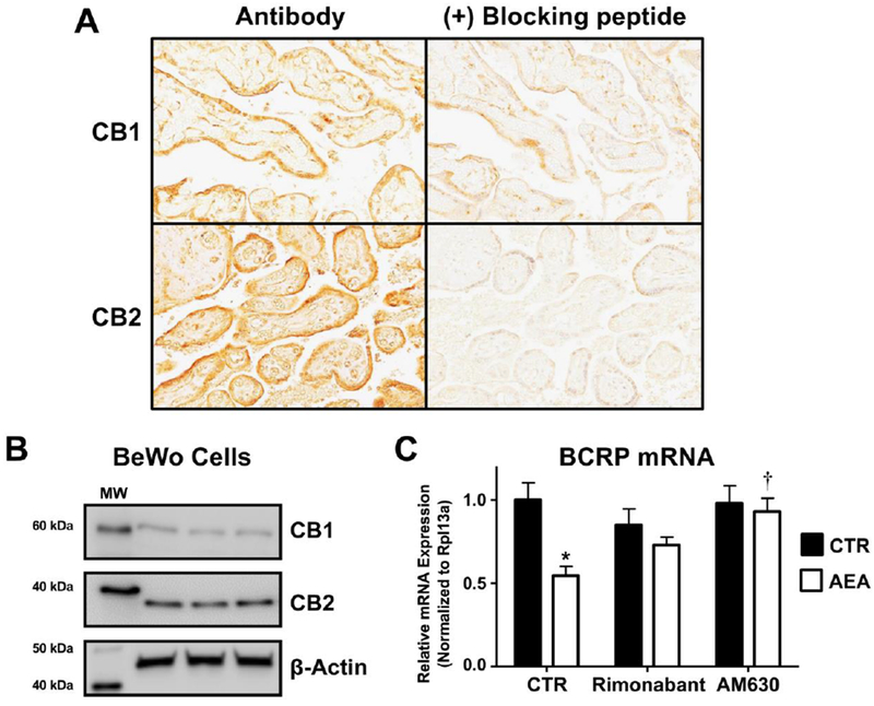
Fig. 5. Expression and function of cannabinoid receptors in human term placentas and human BeWo trophoblast cells.

A. Immunohistochemical staining of the CB1 and CB2 receptors in term human placentas. Sections of paraffin-embedded healthy term placentas (5 μm) were prepared and stained with antibodies against CB1 and CB2 as well as respective blocking peptides and visualized using a Vectastain DAB kit (brown staining) as described in the Materials and Methods. Magnification 20×. B. Protein expression of the CB1 and CB2 receptors in BeWo cells. Lysates were prepared from naïve BeWo cells and analyzed for expression of CB1 (60 kDa) and CB2 (38 kDa) by Western blotting. β-Actin (42 kDa) was used as a loading control. C. CB-receptor dependence of AEA down-regulation of BCRP. qPCR was performed on cells treated with vehicle control (CTR), rimonabant (0.1 μM), or AM630 (0.1 μM) with or without AEA (10 μM) for 24 h. Black bars represent vehicle-treated cells and white bars represent AEA-treated cells. Data are presented as mean ± SE (n = 4). Asterisks (*) represent statistically significant differences (p < 0.05) compared to control cells. Daggers (†) represent statistically significant differences (p < 0.05) compared to AEA-treated cells.
