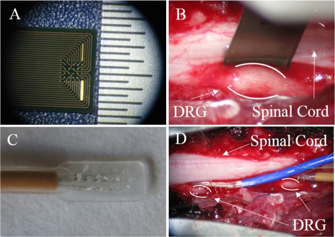
Figure 1.
Images showing non-penetrating microelectrode arrays used in this study. (A) Close-up of 32-channel array manufactured by MCS. Hashes in scale bar separated by 500 µm. (B) MCS array shown placed on the surface of a lumbar DRG, between the DRG and the spinal cord. (C) Close-up of 16-channel array manufactured by PMT. Electrode sites separated by 1 mm. (D) PMT arrays shown placed on the surface of two DRG, between the DRG and the spinal cord.

