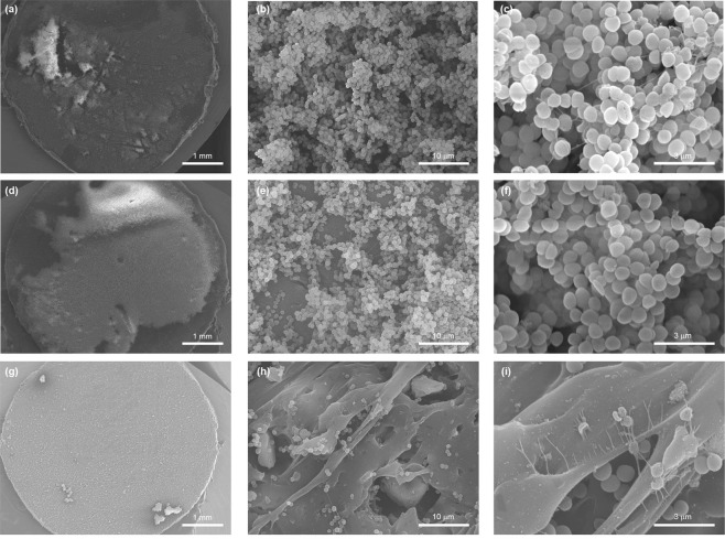
Figure 3.
Scanning electron micrographs of S. epidermidis biofilm (5 days) formed on the surface of untreated TPU (a–c) and TPU composites with 5 wt% (d–f) and 50 wt% (g–i) of glass content. a, d and g panels, show the colonization of polymer surfaces by biofilms (magnification x20). The multilayer biofilm formed on untreated TPU is shown in the panel b (magnification x2.500) and c (magnification x10.000). Water channels and mucoid material appendages implicated in intercellular connection are observed. e and h panels (magnification x 2.500) and f and i panels (magnification x10.000) show the effect of the ZnO content in composites in biofilm architecture and cellular viability.

