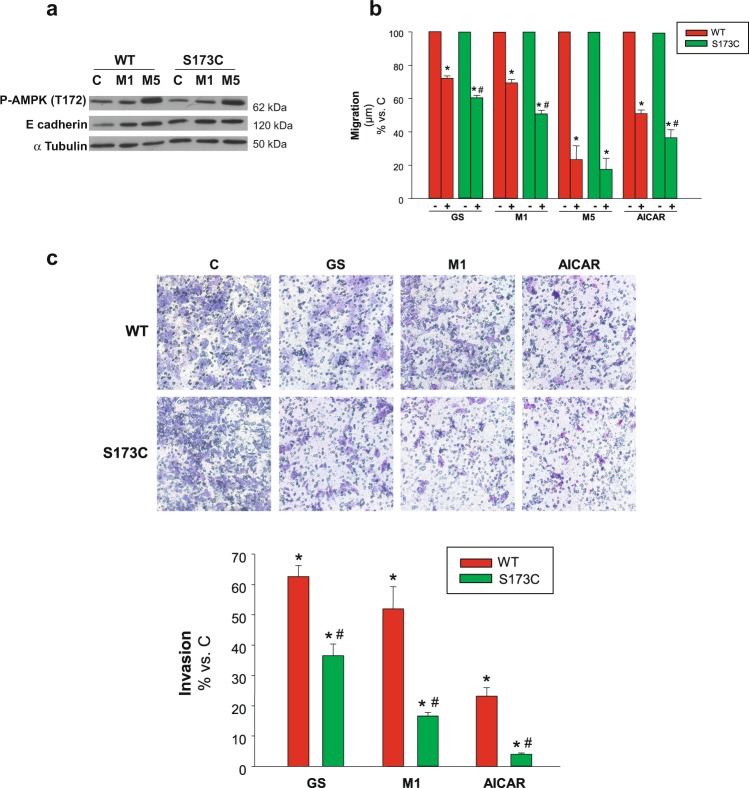
Figure 6.
Contribution of Ser173 regulatory site of AMPKα to cell migration/invasion. (a) C3A cells stably expressing either AMPKα(S173C) (S173C) or wild type AMPKα(S173) (WT) were incubated with complete DMEM alone (C) or in the presence of 1 mM (M1) or 5 mM (M5) metformin for 24 h. P-AMPKα(T172) and E-cadherin protein levels were detected in cell lysates. α Tubulin was used as loading control. Selected lanes for each detection are in their original order and correspond to the same gel, and they are shown after cropping, aligning and separating them by white space. Full-length blots are available in Supplementary Dataset. Immunoblots show an experiment representative of 3 independent experiments (b) C3A cells stably expressing either AMPKα(S173C) (S173C) or wild type AMPKα(S173) (WT) were subjected to scratch wounding (0 h) and incubated in complete DMEM alone (C), or in the presence of 1 mM (M1) or 5 mM metformin (M5), or 1 mM AICAR (AICAR), or glucose starved by incubation in no-glucose DMEM (GS) for 24 h. Data were obtained as indicated in Fig. 1d, and values are expressed as percentages of C. (c) S173C and WT cells were plated in transwell chambers as detailed in Fig. 2 and incubated with complete DMEM alone (C), or in the presence of 1 mM metformin (M1), or 1 mM AICAR (AICAR), or subjected to glucose starvation (GS) for 48 h, and processed as described in Fig. 2. Values represent the mean ± SEM of 3 experiments. *P < 0.05 vs. C. #P < 0.05 vs. WT.

