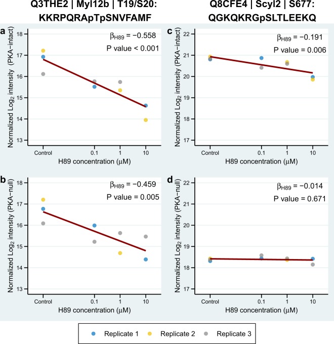
Figure 2.
H89 effects on Myl12b and Scyl2 phosphorylation. Examples of phosphosites from Myl12b and Scyl2 are shown along with their associated linear models. Concentration of H89 (log10 scale) is plotted against log2-transformed intensity normalized for the batch effect. Dark red lines correspond to fitted linear models with slopes equal to βH89 and intercepts averaged from 3 replicates. UniProt accession numbers, gene symbols, phosphorylation positions, and centralized amino acid sequences are shown on the top. Phosphorylation at T19 and S20 in myosin regulatory light chain (Myl12b) are seen to be significantly decreased by H89 treatment in both PKA-intact (a) and PKA-null cells (b). Phosphorylation at S677 in SCY1-like protein 2 (Scly2) is inhibited by H89 in PKA-intact cells (c), but not in PKA-null cells (d), suggesting that this phosphosite is a PKA target.

