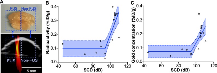
Figure 6.
(A) The ex vivo mouse brains were sliced coronally into 2-mm slices, and the slice containing the targeted brainstem was cut into two halves for quantifying the radioactivity in each half (illustrated by the rectangle boxes). The SCD was also averaged within each half of the brain for identifying the correlation between SCD and radioactivity. (B) The radioactivity of 64Cu-AuNCs quantified using gamma counting and (C) the gold concentration of 64Cu-AuNCs quantified using ICP-MS in the FUS-treated halves and contralateral non-treated halves were well correlated with the corresponding spatial-averaged SCD within the same brain regions using the segmented linear regression (R2 = 0.62 for gamma counting and R2 = 0.53 for ICP-MS).

