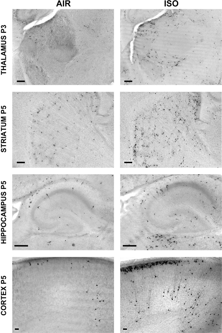
Figure 1.
Age at greatest vulnerability to increased apoptosis induced by ISO in the developing mouse brain is region-specific. Mouse pups were exposed to 1.5% ISO or AIR only for 3 h on P3, P5, or P7, and processed for AC3+ IHC beginning 6 h after initiation of exposure. Representative AC3+ IHC images of mouse pup brain at the ages at which the greatest increase in apoptotic neurodegenerative response induced by ISO above typical developmental levels (AIR) was observed. Thalamus, P3; Hippocampus, P5; Striatum, P5; Cortex; P5. Scale bar is 50 µm.

