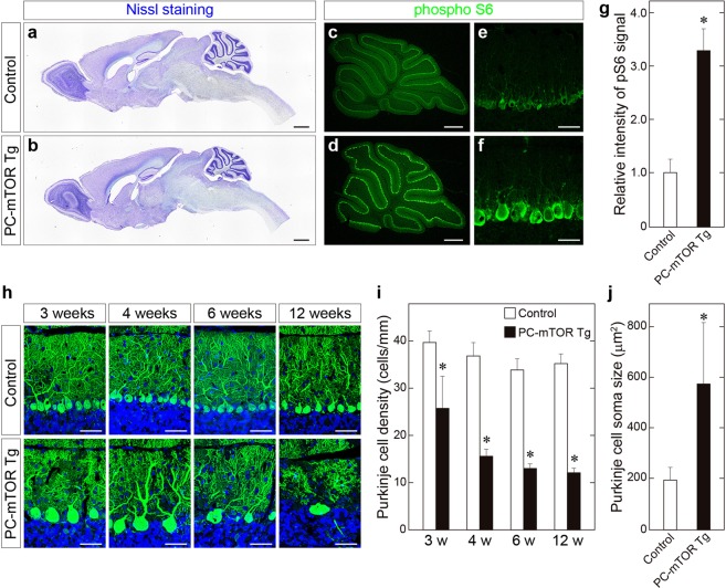
Figure 1.
Activation of mTORC1 signaling in Purkinje cells of PC-mTOR Tg mice. (a,b) Brain morphology of control and PC-mTOR Tg mice at 12 weeks of age. Parasagittal brain sections were stained with cresyl violet. Overall brain morphology was preserved in PC-mTOR Tg mice except the atrophied cerebellum. (c–g) Enhanced phosphorylation of S6 protein in Purkinje cells of PC-mTOR Tg mice. The sagittal sections from control (c and e) and PC-mTOR Tg (d and f) cerebella at 4 weeks of age were immunostained with an antibody to phosphorylated S6 protein. Immunopositive signals were quantified in panel g. (h–j) Age-dependent changes of cerebellar morphology of control and PC-mTOR Tg mice. In PC-mTOR Tg mice, Purkinje cell density was significantly decreased with age, and hypertrophied Purkinje cells were obvious from 3 weeks of age. The density and soma area of Purkinje cells were quantified in panel i and j, respectively. Scale bars, 1 mm (a and b); 500 μm (c and d); 50 μm (e,f and h). *p < 0.001 by Student t-test (g,i and j); control, n = 12 cells from 2 mice; PC-mTOR Tg, n = 12 cells from 2 mice (g); n = 10 slices from 2 mice at each time point (i); control, n = 101 cells from 2 mice; PC-mTOR Tg, n = 133 cells from 2 mice (j).

