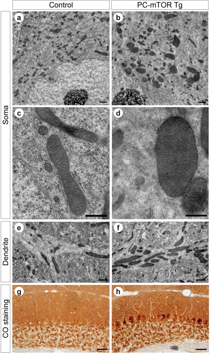
Figure 6.
Abnormal mitochondrial morphology and activity in PC-mTOR Tg mice. (a–f) Electron microscopic images of Purkinje cells of the control and PC-mTOR Tg mice. Enlarged mitochondria were observed in both cell bodies (a and b) and dendrites (e and f). High magnitude images of cell bodies are displayed in c and d, demonstrating that the internal lamellar structure of cristae was preserved even in the enlarged mitochondria in PC-mTOR Tg mice. (g,h) Enhanced mitochondrial respiratory activity in PC-mTOR Tg mice. Enzymatic activity of cytochrome c oxidase was visualized in the cerebellar slices from the control and PC-mTOR Tg mice. Scale bars, 1 μm (a,b,e and f); 500 nm (c and d); 50 μm (g and h).

