Figure 5.
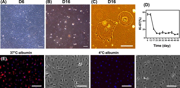
hPSC-derived podocytes exhibit key podocyte phenotypes. IMR90-4 iPSC-derived podocytes were differentiated as illustrated in Fig. 1A. Phase contrast images of IMR90-4 iPSC-derived podocytes were taken at (A) day 6, and (B,C) day 16. (D) Proliferation of cells at different time points was assessed by flow cytometry for Ki67. (E) IMR90-4 iPSC-derived podocytes at day 16 were analyzed with an Albumin Uptake Assay Kit. Alexa Fluor™ 555-labelled albumin is shown in red on a merged DAPI image and the corresponding bright field image is provided on the right. 4 °C was used as a control to prevent endocytosis. Scale bars, 100 µm. Albumin uptake assay was performed three times from three different differentiations.

