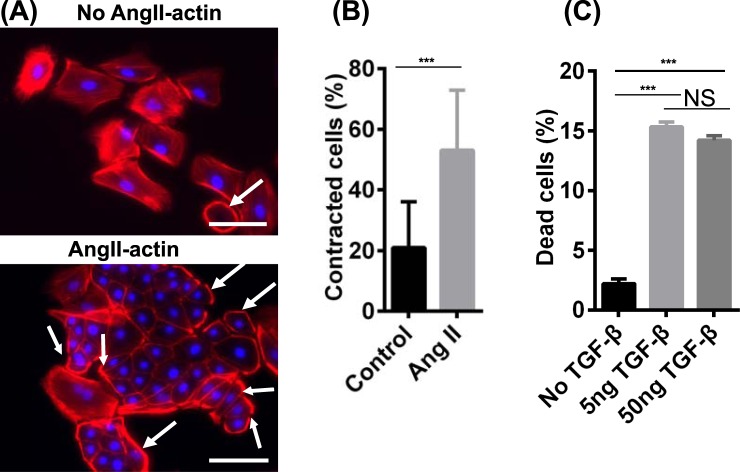
Figure 6.
hPSC-derived podocytes exhibit actin reorganization after angiotensin II treatment and cell death is induced by TGF-β1 treatment. IMR90-4 iPSC-derived podocytes were differentiated as illustrated in Fig. 1A. (A) At day 16, cells were treated with 500 ng/mL Ang II for 6 hr and phalloidin staining was used to assess changes in cytoskeletal structure. White arrows indicate the cells with peripheral F-actin. (B) More than 200 cells over 15 images were analyzed and the percentage of cells with peripherally-organized actin was calculated for control and Ang II-treated cells. Data are presented as mean percentage ± SEM of three independent experiments over 15 images. Scale bars, 100 µm. (C) At day 16, cells were treated with 5 ng/mL and 50 ng/mL TGF-β1for 24 hr. Detached cells were collected from the medium and combined with Accutase-dissociated cells from the substrate. Cells were treated with trypan blue and the numbers of cells incorporating and excluding trypan blue were counted on a hemocytometer. Percentage of dead cells was calculated as the number of cells that incorporated trypan blue divided by the total cell number. Data are presented as mean ± SEM of three independent experiments. Ang II and TGF-β treat experiments were performed three times from three different differentiations.

