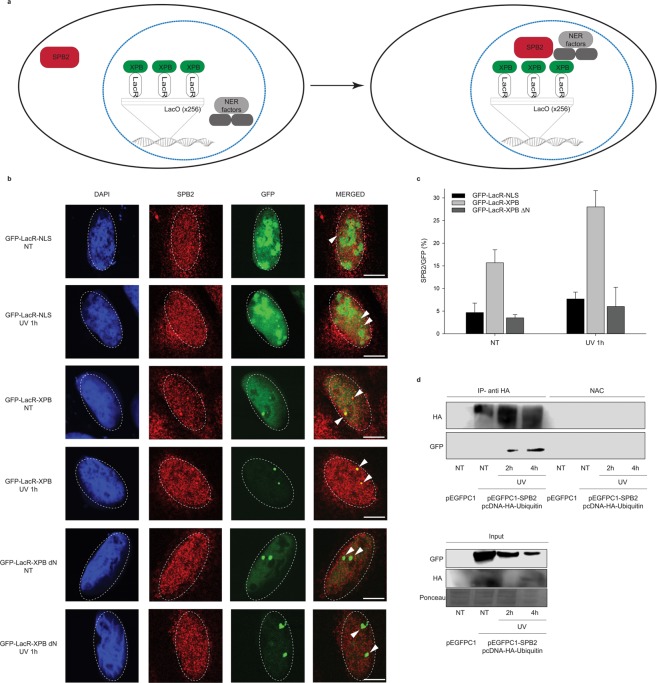
Figure 4.
XPB could anchor SPB2 to the sites of UV damage. (a) Schematic representation of the LacO-tethering assay. (b) Recruitment of SPB2 to the anchored GFP-LacR-NLS or GFP-LacR-XPB or GFP-LacR-XPB∆N is shown in U2OS17 cells. Immunostainings were performed both under normal conditions (NT) and upon UV irradiation (UV 1 h). Arrowheads indicate the cellular position of the LacO arrays. Scale bars represent 5 µm. (c) The values on the graph represent the frequency of the co-localization of SPB2 with GFP obtained from 100 cells in case of each experiment (N = 3). Black columns represent the GFP-LacR-NLS, grey columns represent the GFP-LacR-XPB, while dark grey columns represent the GFP-LacR- XPB∆N data, respectively. (d) Immunoblot detection of SPB2 and ubiquitylated protein interaction in U2OS cells. The success of the immunoprecipitation experiment was controlled with anti-HA antibody and the connection between SPB2 and ubiquitin was detected with anti-GFP antibody (since cells were transfected with plasmid encoding SPB2-EGFP fusion protein) in the precipitated samples both in control and UV treated (2 h and 4 h) samples. Immunoblot detection of GFP and HA signals in the input samples are shown in the lower panel.

