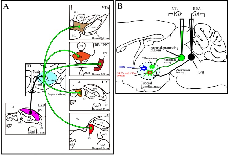
Figure 1.
Schematic diagram of this research. (A) Diagram of axonal projections from LPB to brainstem arousal areas via ORX neurons which were newly identified in this study. (B) Schematic drawings of combined anterograde (black) and retrograde (green) tract-tracing method. Immunohistochemistry was performed on sections of the tuberal hypothalamus (dotted line). CTb negative and ORX positive neurons (ORX+) are represented as blue neurons, CTb positive ORX negative (CTb+) cells are as green neurons and CTb positive ORX positive neurons (ORX+ and CTb+) are as red neurons. LPB; lateral parabrachial nucleus, ORX; orexin, CTb; cholera toxin subunit B, BDA; biotinylated dextranamine, VTA; ventral tegmental area, PPT; pedunculopontine tegmental nucleus, LDT; laterodorsal tegmental area, LC; locus coeruleus, DR; dorsal raphe nucleus.

