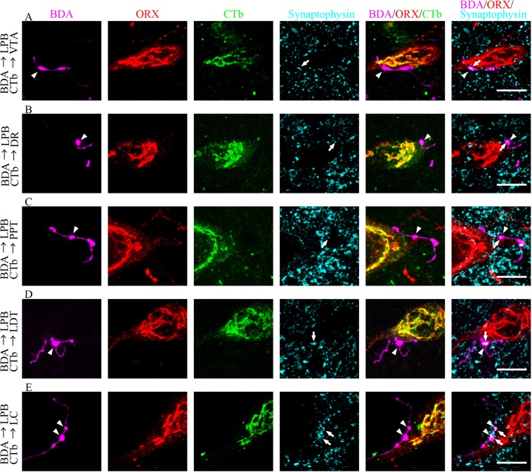
Figure 5.
Confocal microscope images of quadruple fluorescence staining. Images show the appearance of CTb-labeled neurons (green) immunoreactive for ORX (red) and Synaptophysin (cyan) after injection of BDA (magenta) into the LPB and CTb into the VTA (A) DR (B) PPT (C) LDT (D) or LC (E) Two types of merged images, BDA/ORX/CTb and BDA/ORX/synaptophysin, were obtained from the same area. The arrowheads indicate BDA-labeled axons (magenta), which contact to ORX (red) and CTb (green) double-IR neurons. The arrows indicate synaptophysin (cyan) in BDA-labeled axons (magenta), which contact to ORX (red)-IR neurons. Scale bars, 10 μm. LPB: lateral parabrachial nuclei; ORX: orexin; VTA: ventral tegmental area; IR: immunoreactive; CTb: cholera toxin B subunit; BDA: biotinylated dextranamine; PPT: pedunculopontine tegmental nucleus; VTA: ventral tegmental area; DR: dorsal raphe; LC: locus coeruleus.

