Figure 1.
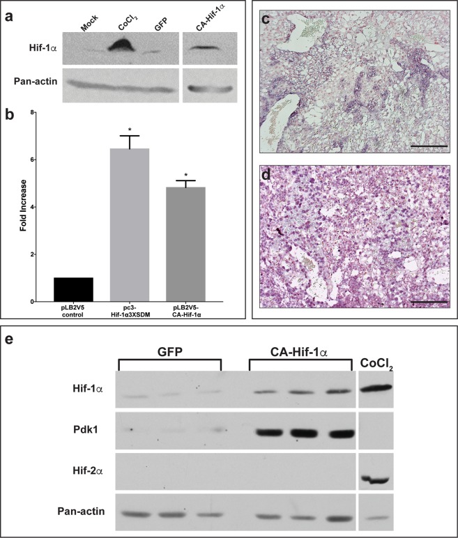
Lentiviral CA-Hif-1α is expressed and activates downstream targets in the mouse placenta following blastocyst infection. (a) Hif-1α protein is stably expressed in COS-7 cells under normoxic conditions following treatment with CoCl2 or transduction with CA-Hif-1α expression construct, compared to non-transduced (Mock) or GFP-transduced controls (raw data, Fig. S2). (b) In luciferase assays using the Pgk1-luciferase reporter, both pc3-Hif-1α3XSDM and the lentiviral pLB2V5-CA-Hif-1α overexpression constructs promoted increased luciferase expression compared to the pLB2V5 control, p < 0.0001 by ANOVA with Tukey’s multiple comparisons test (*). (c,d) Expression of Hif-1α mRNA was detectable by in situ hybridization in E14.5 placentas derived from blastocysts infected with lentiviral particles containing GFP (c) or CA-Hif-1α (d) expression constructs. Expression was more intense and broadly distributed in CA-Hif-1α placentas. (e) CA-Hif-1α placentas collected at E19.5 showed increased expression of Hif-1α protein and its downstream target, Pdk1 compared to GFP controls, but an absence of Hif-2α protein, as detected by Western blot. CoCl2-treated COS7 cells were used as a positive control for Hif-1α and Hif-2α protein expression. Pan-actin was used as a loading control (a,e), (raw data, Figs S3–6) Scale bar = 100 μm.

