Figure 2.
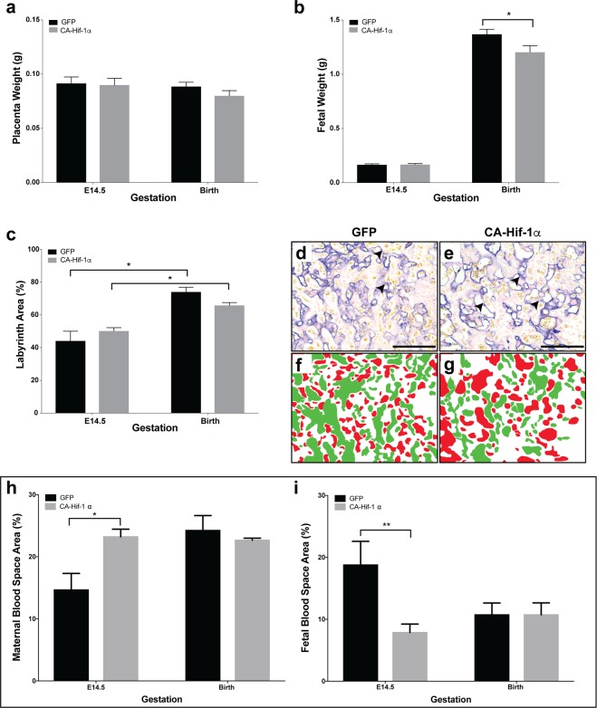
Expression of CA-Hif-1α in trophoblast cells results in reduced fetal weight and altered placental development. (a) Placental weights at E14.5 or birth were not significantly altered by CA-Hif-1α expression, compared to GFP controls. (b) At E14.5, fetal weights from blastocysts infected with CA-Hif-1α were not significantly different, compared to GFP-infected controls (E14.5 GFP pups: n = 7 pups from 3 pregnancies; E14.5 CA-Hif-1α pups: n = 8 from 4 pregnancies). At birth, however, CA-Hif-1α pup weights (n = 21 pups from 6 pregnancies, avg. 1.20 g) were significantly reduced by 12.10%, compared to GFP control pup weights (n = 23 pups from 8 pregnancies, avg. 1.37 g), (*p = 0.03; by mixed model analysis). (c) Assessment of the different layers of the placenta showed that at E14.5 and at birth, the labyrinth layers of CA-Hif-1α and GFP placentas were comparable to each other and that both underwent a statistically significant increase in size between E14.5 and birth (*p = 0.0002; two way ANOVA with Sidak’s Multiple Comparison test). (d,e) Alkaline phosphatase staining in GFP and CA-Hif-1α placentas at E14.5, respectively, is expressed in sinusoidal TGCs and identifies maternal blood spaces in the labyrinth layer (arrowheads). Pseudo-colouring of fetal (green) and maternal (red) blood spaces in GFP (f) and CA-Hif-1α (g) images, which when quantified, identified changes in the makeup of maternal and fetal blood spaces in the labyrinth layer, suggesting reduced branching and limited development of the fetal capillary network. This difference was evident at E14.5 but not at birth (h,i); *p = 0.0255 maternal, **p = 0.0246 fetal by two way ANOVA with Sidak’s Multiple Comparison test. Scale bar = 50 μm.

