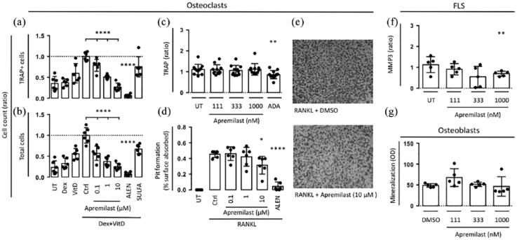
Figure 2.
(a,b) Human bone marrow mononuclear cells (n = 6) were plated and incubated with vitamin D (VitD), dexamethasone (Dex) and compounds for 7 days. TRAP-positive cells and total cells were counted. (c) SFMCs were cultured for 21 days untreated (UT), or treated with DMSO, apremilast or ADA and TRAP (n = 11) secretion was measured. (d) RAW264.7 mouse macrophages were stimulated with RANKL and compounds for 7 days and surface pitting was measured. (e) Representative photographs of osteoclast pit formation. (f) Secretion of MMP3 by FLSs were cultured for 48 h untreated (UT) or treated with DMSO control or apremilast (n = 6). (g) Human osteoblasts were cultured for 14 days untreated (UT) or treated with DMSO control or apremilast and mineralization was assessed (n = 5). Boxes and bars indicate mean and SD. * p < 0.05. ** p < 0.01. **** p < 0.0001.
ADA, adalimumab; DMSO, dimethyl sulfoxide; FLS, fibroblast-like synovial cell; MMP3, matrix metalloproteinase 3; RANKL, receptor activator of nuclear factor kappa B ligand; SD, standard deviation; SFMC, synovial fluid mononuclear cell; TRAP, tartrate-resistant acid phosphatase.

