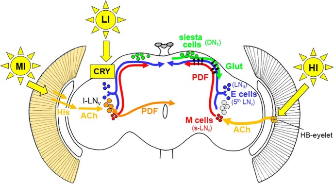
Figure 6.
Model for light input to the circadian clock network of Drosophila. LI light is sensed by CRY in the clock neurons themselves (indicated in the left brain hemisphere) and can quickly reset their oscillations to light (Vinayak et al., 2013). MI light is perceived by the compound eyes, transferred via histamine (His) to the optic lobes and via acetylcholinergic (ACh) interneurons to the large ventrolateral neurons (l-LNv; Muraro and Ceriani, 2015; indicated in the left brain hemisphere). The l-LNv signal via the neuropeptide PDF to the morning (M) and evening (E) cells, respectively (short orange arrow), and to the contralateral hemisphere (long orange arrow). The signaling of HI is shown in the right hemisphere. HI light is perceived by the HB eyelets that signal via ACh to the M cells (s-LNv; Schlichting et al., 2016). The M cells might signal via PDF directly to the E cells (shown by arrows) and inhibit E-cell activity or to the siesta cells (DN1) in the dorsal brain (Guo et al., 2016; Liang et al., 2017). The siesta cells then signal via glutamate (Glut) to the E cells (3 CRY-positive LNd and the fifth sLNv) and delay the onset of E activity (Guo et al., 2016).

