Figure 1.
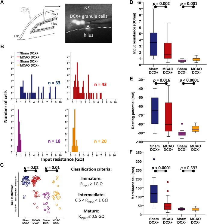
Stroke accelerates cellular maturation of DCX+ ABGCs. A, Schematic illustration of the recording configuration in the granule cell layer of the dentate gyrus. DCX+ phenotype of recorded cells was confirmed by the presence of fluorescence signal in the pipette tip in on-cell configuration (arrowhead). S, Stimulation electrode; mol.l., molecular layer; g.c.l., granule cell layer. B, Histograms of Rinput in the studied population of sham DCX+ (n = 33), MCAO DCX+ (n = 43), sham DCX− (n = 18), and MCAO DCX− (n = 20) cells. The distribution of Rinput in the MCAO DCX+ group is shifted to the left, indicating a more mature phenotype, while in the MCAO DCX− group it is shifted to the right, indicating that stroke has selective effects on different cell subpopulations. C, Distribution of cells within the groups of mature, intermediate, and immature cells (right, definitions) shows a shift toward a more mature phenotype in DCX+ neurons after MCAO, while MCAO DCX− neurons are differently affected. D, Comparative boxplot distribution of Rinput in the four groups shows a significant decrease (more than half) in DCX+ neurons after stroke. E, F, Resting membrane potential (E) and membrane tau (F) also show subpopulation-specific modifications after MCAO, with a maturation shift in DCX+ neurons. See also Table 1 for statistical results.

