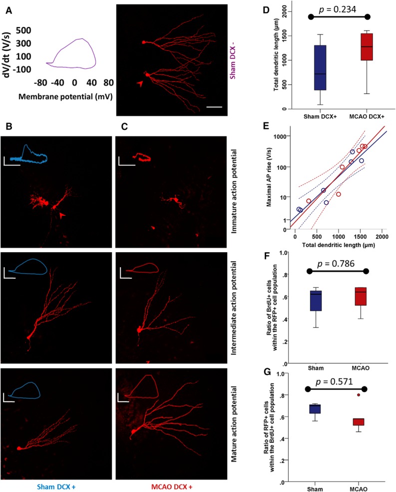
Figure 4.
Conserved morphofunctional coordination in DCX+ neurons after MCAO. A, Right, Biocytin staining appearance of sham DCX− neurons shows an extensive, broad dendritic tree. Left, These cells have fast AP rise velocities. B, C, Morphological maturation follows maturation of intrinsic excitability in both sham DCX+ (B) and MCAO DCX+ (C) neurons (arrowheads indicate cells whose phase plots are represented. Scale bars, 50 μm. Calibration: immature AP, 20 mV, 10 V/s; intermediate AP, 30 mV, 50 V/s; mature AP, 20 mV, 200 V/s. D, Dendritic morphology quantified as TDL by Sholl analysis did not change significantly in DCX+ ABGCs (n = 7) after MCAO (n = 6). E, A high level of correlation between morphological maturation (TDL) and functional maturation (maximal AP rise) was present in sham DCX+ ABGCs and maintained after MCAO. F, The proportion of BrdU+ (born after the insult) cells within the RFP+ cell population (a marker under the control of the DCX− promoter) does not change after MCAO and lies at ∼60% (n = 3 in sham and n = 5 in MCAO groups). G, The proportion of RFP+ cells within the BrdU+ subpopulation does not change, indicating conserved expression dynamics of RFP protein and hence DCX.

