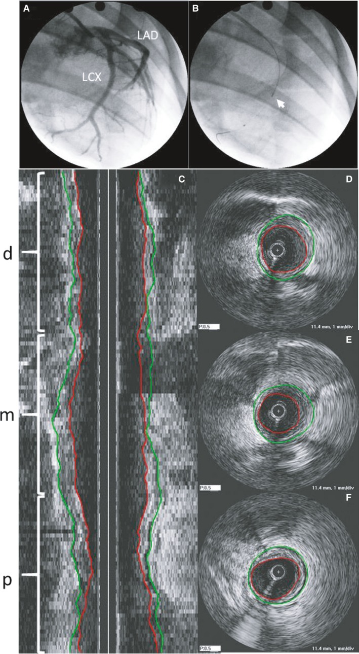
Figure 2.

(A) Typical angiogram showing left circumflex (LCX) and left anterior descending (LAD) and (B) placement of IVUS prior to pullback (white arrow; RAO 60°) in a sedentary animal. (C) Longitudinal cross‐section of IVUS pullback in LCX indicating distal (d), mid (m) and proximal (p) sections with corresponding cross‐sectional images in distal (D), mid (E), and proximal (F). Green lines indicate vessel border (external elastic lamina; EEL) and red lines indicate luminal border.
