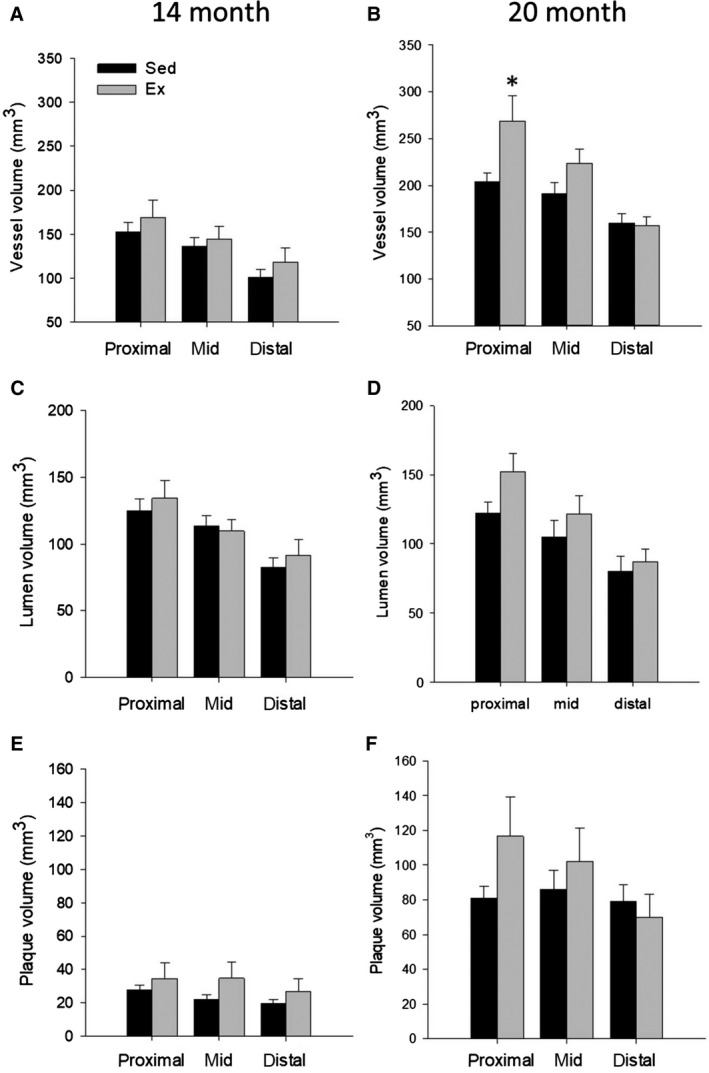
Figure 5.

3D volumetric analysis of the proximal, mid, and distal sections of the left anterior descending artery (LAD) in Sed and Ex at 14 (n = 6 and 8, resp. for all segments) and 20 months (n = 8 and 8, resp. for all segments). There was no significant difference between Sed and Ex at 14 or 20 months for either absolute plaque, vessel, or lumen volume, except a greater vessel volume in the proximal LAD in Ex. *P < 0.05 vs. Sed.
