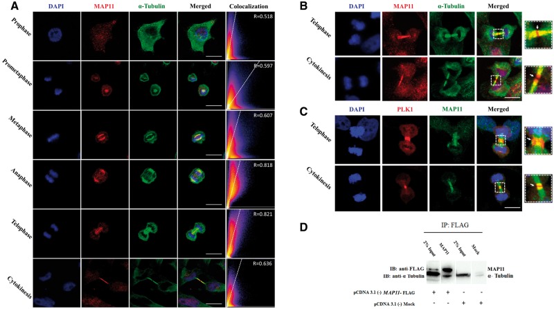
Figure 2.
MAP11 is localized to mitotic spindles and interacts with α-tubulin. (A) Immunofluorescence microscopy of MAP11 in SH-SY5Y cells. Confocal imaging showing endogenous MAP11 co-localizing with α-tubulin during mitosis. Scale bar = 20 µm. Co-localization is highly significant in anaphase and telophase. Graphs in the right panel show Pearson’s correlation coefficient score (denoted by R) between MAP11 and α-tubulin for each image (0 < R < 1; A higher R-score indicates better co-localization between MAP11 and α-tubulin). (B) Immunofluorescence microscopy of MAP11 in SH-SY5Y cells focusing on telophase and cytokinesis. At early cytokinesis, α-tubulin precedes MAP11 in the gap formation followed by cell abscission at the midbody. At late cytokinesis, MAP11 fluorescence projections are sticking out of the microtubule α-tubulin. Right: Enlarged image of the region of interest. White arrow marks the midbody and cell abscission point. Scale bar = 20 µm. (C) MAP11 is co-localized with PLK1 at the edges of each daughter cell’s microtubule extension post cytokinesis abscission. Right: Enlarged image of the region of interest; white arrow marks the midbody and cell abscission point. Scale bar = 20 µm. (D) Co-immunoprecipitation of MAP11 and α-tubulin proteins from SH-SY5Y cells lysates overexpressing MAP11 fused to a FLAG sequence. Immunoprecipitation of cell lysates was performed using anti-FLAG magnetic beads. Immunoblotting was done using primary anti-FLAG and anti-α-tubulin antibodies followed by appropriate secondary HRP-conjugated antibodies.

